Multi-channel MRI Embedding: An EffectiveStrategy for Enhancement of Human Brain WholeTumor Segmentation
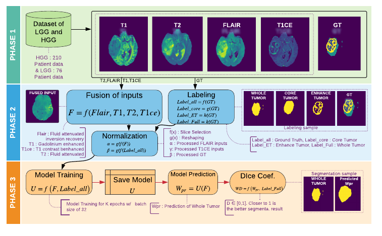
One of the most important tasks in medical image processing is the brain's whole tumor segmentation. It assists in quicker clinical assessment and early detection of brain tumors, which is crucial for lifesaving treatment procedures of patients. Because, brain tumors often can be malignant or benign, if they are detected at an early stage. A brain tumor is a collection or a mass of abnormal cells in the brain. The human skull encloses the brain very rigidly and any growth inside this restricted place can cause severe health issues. The detection of brain tumors requires careful and intricate analysis for surgical planning and treatment. Most physicians employ Magnetic Resonance Imaging (MRI) to diagnose such tumors. A manual diagnosis of the tumors using MRI is known to be time-consuming; approximately, it takes up to eighteen hours per sample. Thus, the automatic segmentation of tumors has become an optimal solution for this problem. Studies have shown that this technique provides better accuracy and it is faster than manual analysis resulting in patients receiving the treatment at the right time. Our research introduces an efficient strategy called Multi-channel MRI embedding to improve the result of deep learning-based tumor segmentation. The experimental analysis on the Brats-2019 dataset wrt the U-Net encoder-decoder (EnDec) model shows significant improvement. The embedding strategy surmounts the state-of-the-art approaches with an improvement of 2% without any timing overheads.
PDF Abstract

