Visualizing Convolutional Networks for MRI-based Diagnosis of Alzheimer's Disease
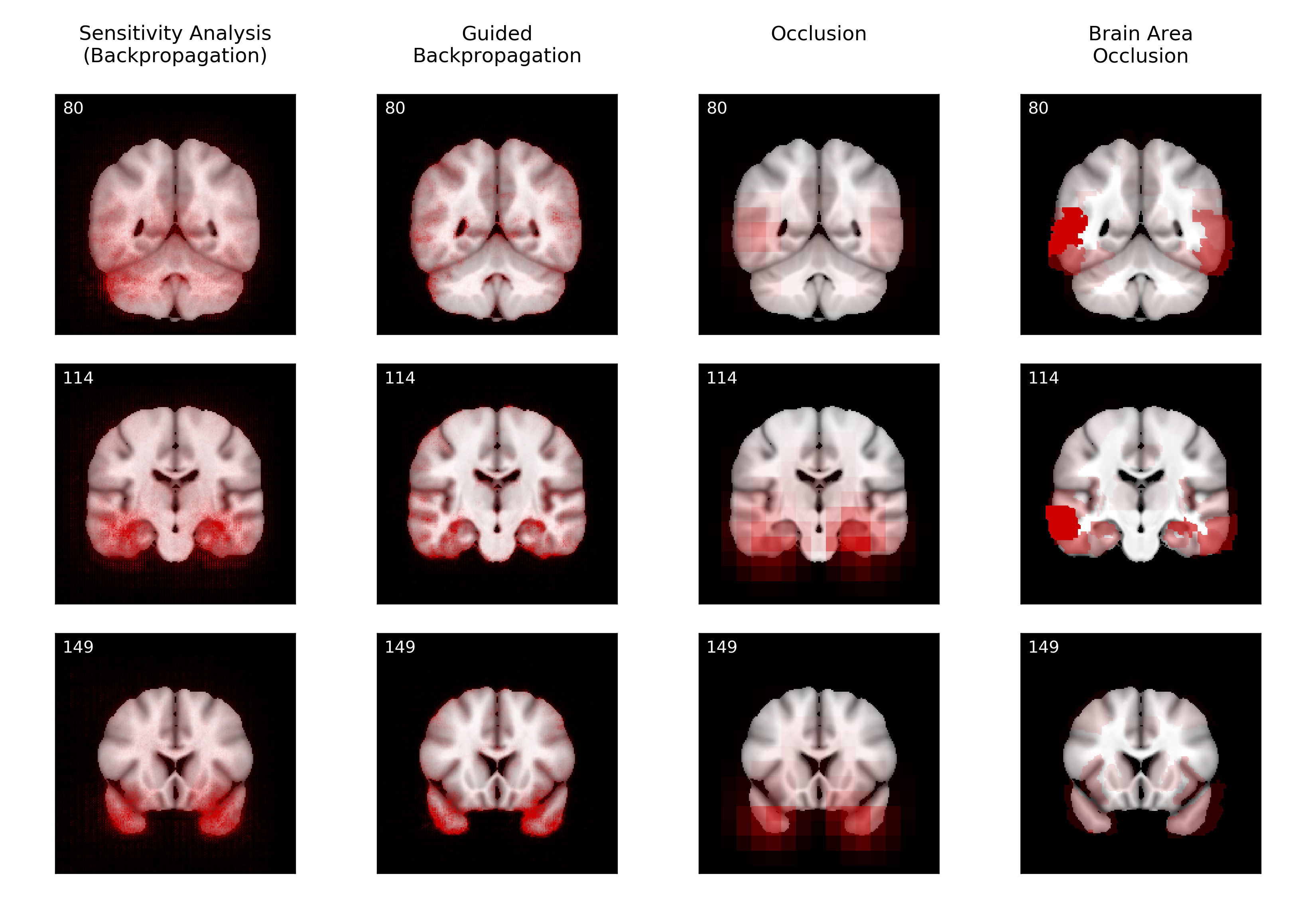
Visualizing and interpreting convolutional neural networks (CNNs) is an important task to increase trust in automatic medical decision making systems. In this study, we train a 3D CNN to detect Alzheimer's disease based on structural MRI scans of the brain. Then, we apply four different gradient-based and occlusion-based visualization methods that explain the network's classification decisions by highlighting relevant areas in the input image. We compare the methods qualitatively and quantitatively. We find that all four methods focus on brain regions known to be involved in Alzheimer's disease, such as inferior and middle temporal gyrus. While the occlusion-based methods focus more on specific regions, the gradient-based methods pick up distributed relevance patterns. Additionally, we find that the distribution of relevance varies across patients, with some having a stronger focus on the temporal lobe, whereas for others more cortical areas are relevant. In summary, we show that applying different visualization methods is important to understand the decisions of a CNN, a step that is crucial to increase clinical impact and trust in computer-based decision support systems.
PDF Abstract

